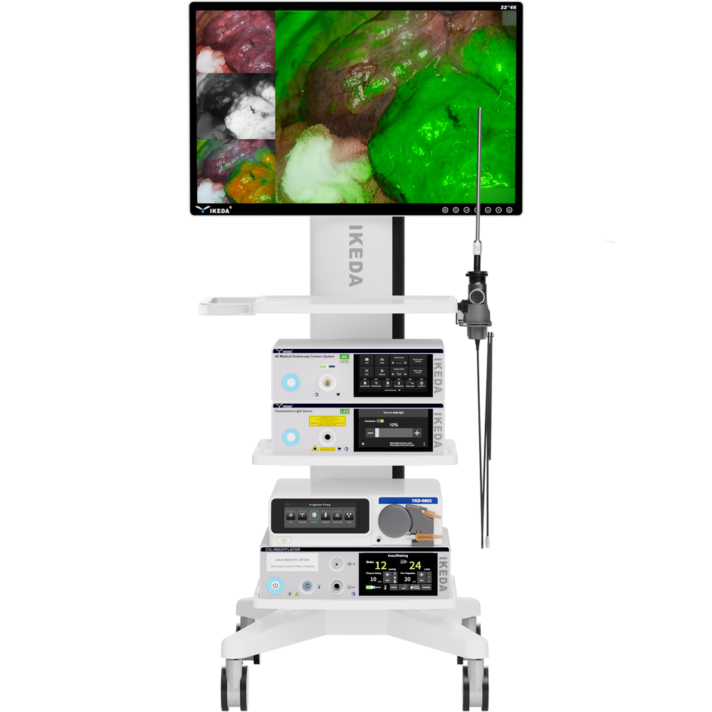
4K ICG/NIR fluorescence endoscope camera system is an optical imaging system that can simultaneously absorb visible light and near-infrared fluorescence.
The system is used with the fluorescent contrast agent indocyanine green (ICG) which generates and emits fluorescence with near-infrared excitation light.
The highly sensitive 4K camera can capture visible light and near-infrared fluorescence, and show image on monitor.
It adopts an imaging solution that integrates 4K and fluorescence technology, with dual-channel 4K ultra-high-definition imaging and extremely precise fluorescence navigation.
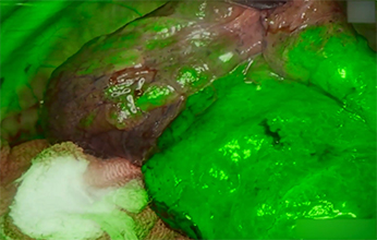
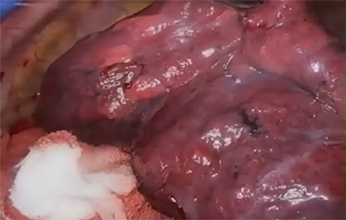
The fluorescence endoscope camera system is a high-end medical imaging device that integrates visible light imaging and fluorescence imaging. Through near-infrared fluorescence (NIR) or ICG labeling technology, it can display tissue blood flow, lymphatic distribution or tumor boundaries in real time, significantly improving surgical accuracy and safety.
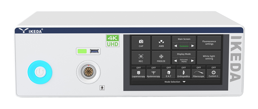
• Pre-set surgical scenarios with one-touch switching
• Built-in 4K video storage
• One-touch full-screen/split-screen toggle
• 7-inch smart LCD touchscreen
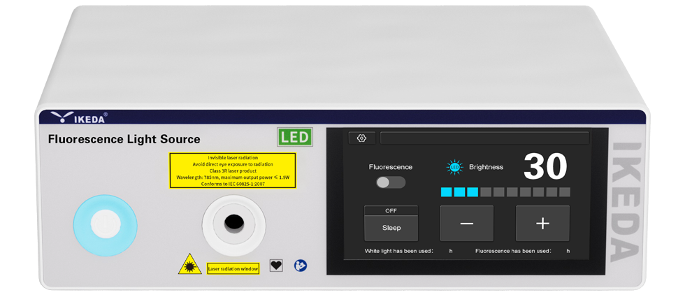
• 20-level brightness adjustment One-touch fluorescence/white mode toggle & sleep mode
• Service life ≥30,000 hours
• Color temperature range: 3,000-7,000K
• 7-inch smart LCD touchscreen
• One-touch white balance:AWB button for automatic white balance calibration
• Multifunction button:Customizable button functions per surgeon preference
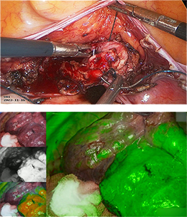
• Four display modes:White light, Fluorescence, Fusion, Grayscale gradient
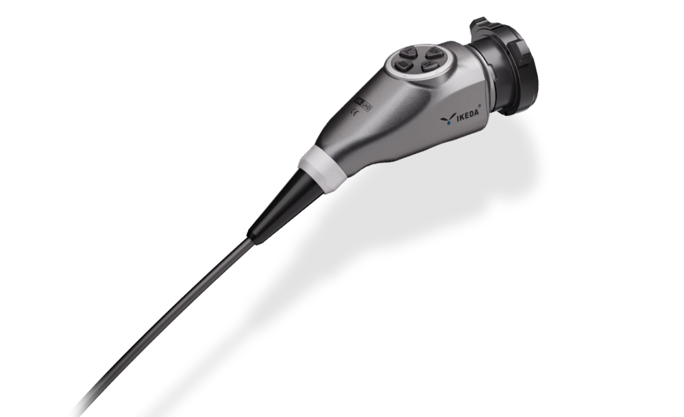
• IPX8 waterproof/Ergonomic design
• Ultra-high resolution compatible with 4K imaging systems
• Autoclavable & plasma sterilization compatible
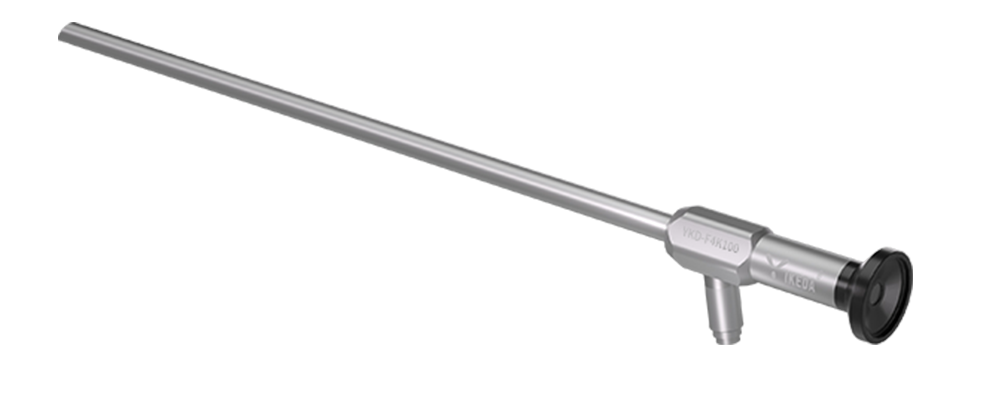
• Full medical-grade stainless steel endoscope body
• Sapphire protective window with scratch-resistant coating
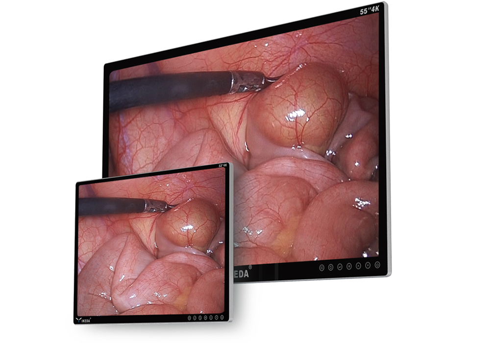
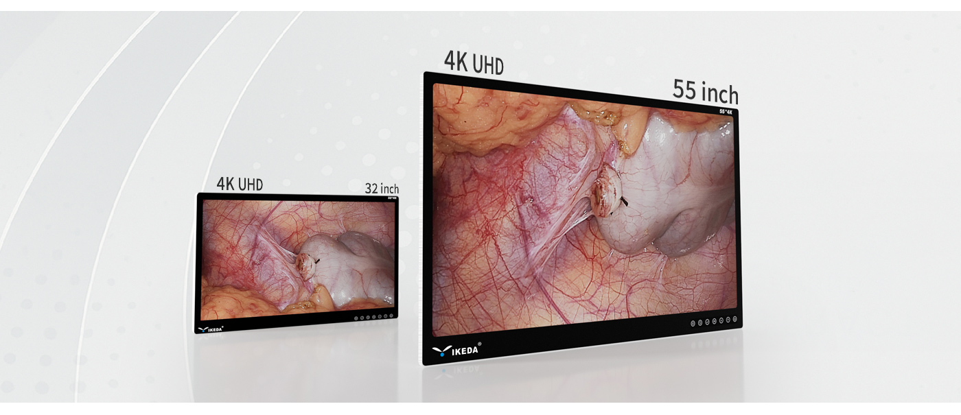
• 3840x2160 ultra-high-definition image output, showing richer textures and more accurate tissue identification
• 3840x2160 pixel image output, showing richer texture, more accurate tissue identification
• Wider color gamut, truly restore the color of organs and tissues, and locate blood vessels, lymph nodes and nerves faster
4K Ultra-HD with Exquisite Image Quality
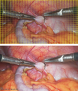
With 4K UHD resolution (3840×2160), the system provides clearer observation of microvasculature and pathological tissue contours, enabling better differentiation of tissue boundaries (e.g., fat, nerves, and vascular junctions).
Breakthroughs in fluorescence technology significantly improve detection sensitivity and imaging stability,
enabling more accurate navigation.
High Fluorescence Sensitivity & Stability
The 4K fluorescence endoscope, when paired with ICG contrast, delivers high sensitivity and precise targeting while accurately displaying tracer distribution with minimal signal loss from distance or angles, ensuring enhanced fluorescence stability.
Smart Algorithm Optimization & Scenario Adaptation
Intelligent imaging algorithms ensure clear surgical visualization with vascular enhancement, thereby improving surgical efficiency and safety. The system adapts to various surgical scenarios.
Pixel-Level Fluorescence Fusion
The fusion of 4K white-light and fluorescence imaging provides surgeons with more accurate fluorescence-assisted positioning.
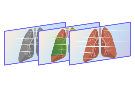
4K + Fluorescence Imaging, Switch Freely Between 4 Imaging Modes
Fluorescence Mode
White Light Mode
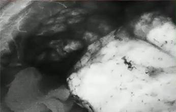
Black and White Fluorescence Mode
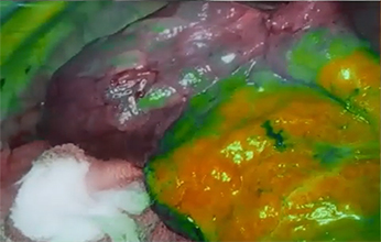
Fluorescence Fusion Imaging Mode
Clinical Departments for ICG Application
 Thoracic Surgery
•Positive Dye Marking for Pulmonary Segmentectomy
•Negative Dye Marking for Pulmonary Segmentectomy
•Fluorescence Imaging of Small Pulmonary Nodules Blood
•Supply Assessment of Gastric Conduit Lymphatic Mapping in Esophageal Cancer
 Gastrointestinal Surgery
•Blood Supply Assessment in Colorectal Surgery Lymph
•Node Mapping in Rectal Cancer Surgery Lymphatic
•Imaging in Gastric Cancer Surgery Tumor Localization in
•Early Gastric Cancer Surgery
 Gynecology
•Pelvic Lymph Node Dissection for Endometrial Cancer
•Inguinal Lymph Node Dissection in Vulvar Cancer Pelvic
•Lymph Node Dissection for Cervical Cancer Ureteral Imaging
 Urology
•Retroperitoneal Lymph Node Dissection (RPLND) for Testicular Cancer
•Sentinel Lymph Node Biopsy (SLNB) and Inguinal Lymph Node Dissection in Penile Cancer
•Bilateral Inguinal, Vascular, and Obturator Nerve Lymph Node Dissection
•Tumor Margin Identification in Partial Nephrectomy
•Lymph Node Dissection in Bladder/Prostate Cancer
Gastrointestinal Surgery
•Demarcation of Right/Left Hemiliver Boundaries
•Intraoperative Tumor Margin Delineation
•Post-hepatectomy Bile Leakage Detection
•Preliminary Assessment of Hepatocellular Carcinoma Differentiation
•Detection of Residual Tumor/Microlesions at Hepatic Resection Margins
•Ultrasound-Guided Portal Vein ICG Fluorescence Imaging for Hepatic Segment Delineation
•Evaluation of Vascular Patency and Graft Function Post-liver Transplantation
Please leave a message here and leave your contact information,
and we will reply to you as soon as possible.